Holistic Approach to Glycemic control

HOLISTIC APPROACH TO GLYCEMIC CONTROL

News - Mar. 30, 2011

Ralph A. DeFronzo, M.D., Professor of Medicine, Chief, Diabetes Division, University of Texas Health Science Center, San Antonio, TX

Introduction 

Ten years ago, our concept of the pathogenesis of type 2 diabetes was quite simple. We recognised that there was underlying moderate-severe insulin resistance affecting muscle and the liver, and this, coupled with beta cell failure, formed the classical triumvirate (1). The insulin resistance in muscle primarily was responsible for the excessive postprandially rise in plasma glucose concentration, while insulin resistance in the liver, in combination with accelerated gluconeogenesis, resulted in an excessive rate of hepatic glucose production which led to an increase in the fasting plasma glucose concentration (2-4). In response to the insulin resistance, the beta cell initially is able to compensate, by appropriately increasing its secretion of insulin to maintain normal glucose homeostasis (1,5,6). However, with time declining beta cell function results first in postprandial hyperglycaemia and subsequently a rise in the fasting plasma glucose concentration and ultimately the onset of frank diabetes (5,7-9). The relative contributions of beta cell failure and insulin resistance to the diabetic state vary between different ethnic populations (10). However diabetes does not become manifest until beta cell failure leads to a failure to compensate for the insulin resistance(1,11).

Natural History of Diabetes

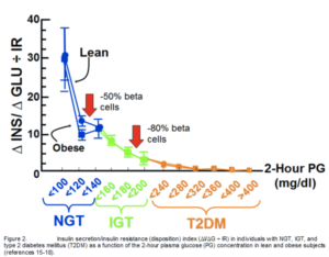
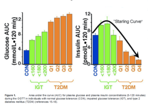
Insulin resistance can be demonstrated long before the onset of T2DM, even before the onset of impaired glucose tolerance (IGT). Thus, in a study of normoglycaemic offspring of two type 2 diabetic parents, it was demonstrated that the insulin resistance already was maximally/near maximally manifest (12-14). These offspring maintained normal glucose tolerance (NGT), because their beta cells were able to recognise the severity of the insulin resistance and appropriately increase the amount of insulin secreted in order to maintain normal glucose homeostasis. However, with advancing age, the beta cells no longer can maintain their high level of insulin secretion and the beta cells begin to fail. With the decline in insulin secretion, the insulin resistance in the liver becomes manifest as a rise in hepatic glucose production during the sleeping hours and results in an elevated fasting glucose (FPG), while in the muscle, following a meal, there is an excessive rise in the postprandial plasma glucose (PPG) concentration. Over the last five years we've learned that the beta cell failure begins much earlier in the natural history of T2DM and is much more severe than previously understood. In the San Antonio Metabolism Study (SAM) and the VAGES Study beta cell function was evaluated in 259 IGT, 201 T2DM and 318 NGT subjects, who were classified as being obese or non-obese, using 2 different cut-points: BMI ≥ 30 kg/m2 or ≥27.5 kg/m2 (15-18). Notably, the value used for the cut-point of obesity was found to make no difference on the results, with the beta cells of both obese and non-obese subjects behaving in the same way. All subjects received a 75 gram OGTT with measurement of the plasma insulin concentrations every 15 minutes to quantitate insulin secretion. On a separate day subjects received a euglycemic insulin clamp to measure whole body insulin-mediated glucose disposal. IGT subjects were divided into tertiles based upon the 2-hour plasma glucose concentration (140-159, 160-179, 180-199 mg/dl). Within the IGT range, insulin secretion initially rose in response to increased plasma glucose levels, but in the upper tertile of IGT, insulin secretion began to decrease, and declined further and progressively through the four quartiles of T2DM. Graphically, this produces an “Inverted-U” shaped curve, similar to Starling’s curve for cardiac function and colloquially might be termed “Starling’s curve of the beta cell” (1) However, insulin levels are not a good reflection of beta cell function, and it's important to distinguish between the plasma insulin response and the health of the beta cell, or beta cell function. Since the beta cell responds to an increment in glucose with an increment in insulin, a better index of its function is to divide incremental insulin response by the incremental glucose response during the OGTT. However, the beta cell also recognises the severity of the insulin resistance and adjusts its secretion of insulin to appropriately match the severity of insulin resistance (6,19). Hence, the gold standard index for beta cell function is the increment in insulin per increment in glucose divided by the severity of insulin resistance, termed the insulin secretion/insulin resistance (disposition) index (I/G÷IR) (15,18,20).

Using the gold standard IS/IR index, across the tertiles of NGT (2-hour glucose during the OGTT: <100, 100-119, 120-139 mg/dl) beta cell function progressively declined resulting in a loss of 50-60% by the third tertile (15-18). This progressive decline in beta cell function continues throughout the range of dysglycaemia and in the upper tertile of IGT, by which time individuals are maximally/near maximally insulin resistant, beta cell function has decreased by 75-80%.


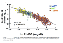
Such patients, while not classified as “diabetic” by current definitions, are clearly so from the pathophysiological standpoint. It takes very little further decline in beta cell function for subjects in the upper tertile of IGT to progress to overt T2DM with markedly elevated plasma glucose levels (FIGURE 2). The severity of this beta cell dysfunction in IGT individuals is not fully understood by most physicians. These results clearly indicate that adiabetic treatment needs to be initiated much eathan currently ordained, in order to protect the remaining 20% of beta cell function. The arbitrary nature of current diagnostic criteria is exemplified by the log transformation of these data.

When the In of the IS/IR index (measure of glucose tolerance) in NGT/IGT/T2DM subjects, there is a strong linear relationship, with no obvious diagnostic cutpoints (21) (fig33). The need for earlier intervention, at the stage of IGT, is further reinforced by the observation that, IGT patients with a HbA1c of ~6%, up to 10% of individuals already have background diabetic terinopathy 922,23) and pheripheral neuropathy is common (24). As afurther confounder to didactic treatment algorithms, individuals are known to differ in their genetic susceptibility to an elevation in the plasma glucose concentration. Thus many type 2 diabetic individuals have a HbA1c in the 7.5-8.0% range yet they do not develop microvascular complications, while other diabetic patients with HbA1c values of 6.5-7.5% exhibit evidence of diabetic retinopathy and other microvascular complications. This stresses the need to individualize a patient's treatment based upon physical findings indicating the presence of end organ damage.

Treatment Options

Although no drugs currently are approved for treatment of “prediabetes”, early treatment of IFG/IGT patients should be considered. Such treatments should target the pathogenic mechanisknown to promote beta cell failure and insulin resistance, rather than the classical goal of simply reducing the HbA1c.

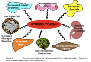
Increased knowledge of the nature of diabetes now recognises that instead of the 3 classic pathologies of the "triumvirate" (1), there is an octet (fig 4) of recognised pathophysiologic disturbances (21) including: (1) adipose tissue insulin resistance characterized by accelerated lipolysis, (2) decreased incretin effect, (3) impaired insulin secredtion, (4) increased glucagon secretion by the alpha cell, (5) hepatic insulin resistantance and accelerated hepatic gluconeogenesis, (6) increased renal glucose reabsorption, (7) muscle insulin resistance and (8) brain insulin resistance to the appetite suppressant effect of hyperinsulinemia (25,26). The sheer number of these pathophysiologic disturbances dictates the ned for multiple drugs used in cmbination to effectively reduce the plasma glucose concentration inT2DM patients since no single antidiabetic agent can address all of these abnormalities.

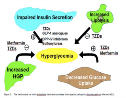
Not only will early intervention serve to preserve the remaining beta cells, but will help attenuate other irreversible damage, e.g. the microvascular and macrovascualr complications that have already begun in the prediabetic state. The mechanisms via which antidiabetic medicationaddress these specific pathogenic disturbances are shown in (FIGURE 5):
  1. Hepatic Insulin Resistance/Increased HGP:Metformin and thiazolidinediones (TZDs) are potent insulin sensitizers in the liver, beiadditive and complimentarysince TZDs act via the classical insulin signalling pathway, while metformin's action is mediated via the AMPK-stimulated pathway (27,28). Neither metformin nor the TZDs carry the risk of hypoglycemia and both have been shown to give some protection against cardiovascular disease (29,30). Metformin also blunts the weight gain associated with TZDs.
  2. Muscle Insulin Resistance: Metformin is ainsulin sensitizer, while TZDs are powerfu
  3. Adipocyte Insulin Resistance and AccelerateLipolysis: TZDs are the only currently availablanti-diabetic drugs that improve adipocyte insulin resistance, inhibit lipolysis, reduce the excess production of free fatty acids (FFAs), and decrease the plasma FFA concentration (32-34).
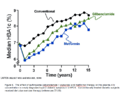
  4. Beta Cell Dysfunction: TZDs have the greatest amount of data demonstrating their durable efficacy and preservation of beta cell function (reviewed in reference #21) while the GLP-1 analogue exenatide now has 3.5 year durability data (35). There currently are only short term data regarding the effect of the DPP4 inhibitions (36) to preserve beta cell function, and they inherently are less potent than the GLP-1 analogues. Sulphonylureas (SUs) initially stimulate insulin secretion and the increased plasma insulin levels overcome the insulin resistance and reduce plasma glucose level. However, within 1-1.5 years the SUs consistently fail because they do not stop the progressive beta cell failure that is characteristic of the diabetic state (FIGURE 6).

Lifestyle, Sulphonylureas and metformin

UKPDS demonstrated that conventional therapy with diet plus excercise failed to prevent the long -term rise in HbA1c that occured secondary to progressive beta cell failure (37). UKPDS also documented that monotherapy with a sulfonylurea glibenclamide) or with metformin caused an initial drop in HbA1c, but the effect was nt durable and the HbA1c had returned to or above baseline values by year 5 (fig 6) (37-39). Because of the progressive rise in HbA1c with monotherapy, diabetic subjects on a SU had metformin added, while subjects on metformin had a SU added. After a brief drop with addition of the second agent, HbA1c levels rose once again, demonstrating that durability is not achieved even when metformin/SU therapies are used in combination (37-43). despite the prgressive rise in HbA1c, a 1% difference in HbA1c between the conventional and '"intensive" therapy arms in UKPDS was maintained throughout the study and this was associated with a 37% reduction in eye, kidney and nerve disease, providing definitive proof in T2DM that improved glycaemic control provides protection against the microvascular complications (44).

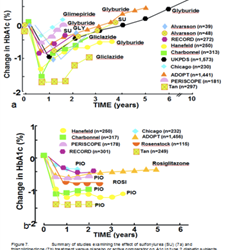
However, it is clear that even combined metformin/SU therapy failed to achieve durable glycemic control and after 15 years the HbA1c had risen to ~8.5% despite the fact that ~65% of the diabetic patients also were on insulin therapy. The results have been replicated by all comparator studies with SUs in which the study duration was 1.5 year in duration (FIGURE 7a), reinforcing the facthat SUs do not provide durable glycemic control. In contrast, 8 large studies, lasting up to 5 years have demonstrated the durability of HbA1c control (fig 7b)


Thiazolidinediones (TZDs)

TZDs have 2 major mechanisms of action to improve glycemic control

  1. They are potent insulin sensitizers in muscle, liver, and adipocyes (27,28,31-33)
  2. They preserve beta cell function (44,45)
TZDs bind to the PPAR y receptor in muscle/liver and beta cells, activating genes that augment insulin action, promote beta cell health, and inhibit beta cell apoptosis. TZDs also reverse lipotoxicity (46). Thus, TZDs bind to the PPARy receptor in subcutaneous adipose tissue causing them to divide. The net result is an increase in the number of small fat cells. In addition, the TZDs activate all of the genes involved in lipogenesis in these newly formed small fat cells (46-48) leading to a reduction in plasma FFA concentration and decreased flux of FFA into muscle, liver, and beta cells. In addition, the TZDs activate PGC-1 which is the master gene that regulates mitochondrial biogenesis (49-51). The increase in mitochondral gene expression leads to enhanced intracellular lipid oxidation and a reduction in toxic lipid metabolites (ceramides, fatty acyl CoAs and diacylglycerol) that cause beta cell apoptosis and insulin resistance 33,45,49,52).
Thus the TZDs address an underlying pathology that all patients with diabetes, including lean T2DM individuals, have: namely, high levels of fat in muscle, liver and beta cell. It also is noteworthy that FFA are a major component atherosclerotic plaques and these FFA are verinflammatory and likely to play an important role in the atherosclerotic process (53-55). Euglycaemic insulin clamp studies performed after 6 months of treatment with pioglitazone have demonstrated a halving of the insulin resistance, accompanied bydecreases in plasma FFA, total muscle fat contentand muscle long chain FACoAs. Since TZDs are powerful insulin sensitizers and improve beta cell function, their potential use in impaired glucose tolerance (IGT) has been investigated. In 4 studies to date, TZDs consistently have decreased the progression of IGT to T2DM: TRIPOD (-52%), PIPOD (-62%), DREAM (-62%) and ACT NOW (-72%). (56-59).


GLP-1 ANALOGUES

GLP-1 analogues are effective in causing a durable reduction in HbA1c, with an equivalent potency to that of the TZDs (56-69). The GLP-1 analogues preserve beta cell function (34,61,62), the latter being key to halting the progressive rise in HbA1c that is characteristic of T2DM (63). The durable effectof GLP-1 analogues to reduce HbA1c has been demonstrated in a 156 week trial with exenatide Exenatide and liraglutide also have a good safety profile and a further advantage over SUs and insuliis that they do not cause hypoglycaemia. Exenatidand liraglutide also have a potent effect to enhancbeta cell function (34,61,62,64). In a 30 week studsubjects received a meal tolerance test before and exenatide, 10 ug bid (64). Exenatide caused a modest reduction in fasting plasma glucose concentration and markedly reduced the postpranrise in plasma glucose concentration due to enhanced insulin secretion, inhibition of glucagon secretion, and delayed gastric emptying (64,65). Most remarkably, the increment in insulin per increment in glucose increased approximately 10-fold, documenting the potent effect of exenatide to augment and preserve beta cell function (64). In a subsequent study the effecet of exenatide on beta cell function was compared with insulin glargine in metformin-treated T2DM patients using the hyperglycaemic clamp technique (61). Glargine, which has no intrinsic effect on the beta cell, and exenatide (up to 15-20 ug tid) reduced the HbA1c tthe same extent (6.8%) after one year of treatment. The glargine group exhibited a 1.31-fold increase in insulin secretion after 1 year of treatment, while in the exenatide arm insulin secretion increased 3.19-fold over baseline. This 10-fold greater increase in insulin secretion (p<0.0001) is largely attributable to a direct effect of exenatide to improve beta cell function, since simply lowering the plasma glucose concentration with glargine, i.e. removal of glucotoxicity, had only a very modest effect to enhance beta cell function (64). Liraglutide hasshown to have a similar beneficial effect on beta cell function (62,66-68). The pleitropic effects of the GLP-1 analogues: (i) improved beta cell function and enhanced insulin secretion, (ii) inhibition of glucagon secretion, (iii) reduction of hepatic glucose production, (iv) decreased appetite mediated through a CNS action, and (v) delayed gastric emptying, make this class of antidiabetic drugs ideal for the treatment of T2DM patients. In toto, the GLP-1 analogues correct 5 of the 8 known metabolic disturbances that comprise the ominous octet while the TZDs correct 4 of the 8 pathogenic disturbances (i) beta cell dysfunction and (ii-iv) insulin resistance in liver, muscle and adipose tissue. In contrast to these agents, metformin has the sole action of increasing insulin sensitivity in the liver, while SUs fail to correct any of the known pathophysiologic defects. In addition, several studies have suggested that SUs may accelerate the development of atherosclerosis (69, 70), a cause for concern given the elevated CV risk in patients with diabetes. DPP4 inhibitors, as monotherapy or add onto other antidiabetic agents, typically reduce the HbA1c by -0.6% to -0.8% (71-73), overall representing a lower glycaemic efficacy than either the TZDs or GLP-1 analogues.

Current Treatment Algorithms

The current ADA/EASD algorithm (74) advocates initiation of therapy with lifestyle modification (weight loss and exercise) plus metformin, even though metformin addresses only one of the 8 known pathophysiologic abnormalities and does not preserve beta cell function. However, results of the UKPDS suggest that metformin also may have some beneficial effects to prevent atherosclerosis. If diet/exercise plus metformin fail to reduce the HbA1below 7.0%, the algorithm recommends the addition of a SU even though the SUs do not correct any of the known pathophysiologic abnormalities present in T2DM and have been shown not to prevent beta cell failure or cause a durable reduction in HbA1c in the UKPDS. If metformin plus a SU fail to reduce the HbA1c < 7.0%, the algorithm suggests the addition a long acting insulin. Other agents (TZDs, GLP-1 analogues), which clearly have a more durable effectto reduce the HbA1c than the metformin and the SUs(FIGURES 6 and 7), are recommended only after metformin/SUs/basal insulin fail. The deficiencies inthe ADA/EASD algorithm draw attention to the need for a different therapeutic approach which is formulated on evidence-based medicine and known pathophysiologic abnormalities and includes TZDs, exenatide, liraglutide, and the DPP4 inhibitowhich have been left out of the algorithm. Even though the DPP4 inhibitors are less effective than theGLP-1 analogues and the TZDs, they are safe and have some advantages in elderly people and inorientals who seem to respond very well to this class.
Lastly, it should be pointed out that the current therapeutic goal for HbA1c (<7.0%) is higher than the diagnostic goal (HbA1c  6.5%), creating a treatment paradox for physicians.

Pathophysiological-based Algorithm

The author strongly advocates that clinicians should initiate treatment earlier than currently practiced and base drug-choices on their ability to correct known pathophysiologic disturbances. This results in an alternative evidence-based algorithm of lifestyle change in combination with a GLP-1 analogue plus a TZD (pioglitazone) plus metformin ab initio, as first presented at the Banting Award lecture at the ADA in 2008 (21). The TZD/metformin combination improves insulin sensitivity and has an additive effect to reduce glycemia, sincethese antidiabetic agents have different mechanisms of action. TZDs and metformin also provide socardiovascular protection and do not run the risk of hypoglycaemia. GLP-1/TZD co-administration also has an additive and complimentary effect to improve glycemia and both drugs have been demonstrated to preserve beta cell function. Moreover, the GLP-1 analogues promote weight loss, offsetting any weight gain associated with TZD use, and the GLP-1 analogues, like the TZDs. do not cause hypoglycemia. In newly diagnosed T2DM patients, the author recommends  a HbA1c goal of < 6.5, and ideally < 6.0%. Since neither the TZDs nor metforminnor the GLP-1 analogues cause hypoglycemia, these drugs can be titrated up yo achieve the HbA1c goal of <
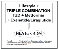
6.0 without hypoglycemia, which is the major obstacle to achieving ootimal glycemic control. Current practice at the Texas Diabetes Institute (SaAntonio, Texas) is to prescribe low-dose (15-30mg/day) pioglitazone to IGT patients with a HbA1c ~6%, while low dose pioglitazone plus low-dose metformin (1000-1500 mg/day) is used for diabetic patients with HbA1c ~7%. In T2DM patients with a HbA1c ≥7.5% at diagnosis, we employ a triple therapy regimen with pioglitazone, metformin and exenatide or liraglutide, thus addresseing the underlying pathophysiology. This approach gets the majority of diabetic patients to the HbA1c goal of ≤ 6.0% (FIGURE 8) and corrects many of the cardiovascular risk factors that are present in T2DM patients.

Summary

We now recognise that diabetes has a complex and multifactorial pathogenesis, with 8 well established pathophysiologic defects. Yet current treatment algorithms focus solely on the control of hyperglycaemia and not on correction of the underlying pathophysiologic disturbances. The primary drugs, metformin and SUs, recommended bythe ADA/EASD do not have a durabale effect either on glycaemic control or prevention of beta cell failure and disease progression, because they fail to treat the core defects present in T2DM patients.
Furthermore, the cut-points for the diagnosis of diabetes are rather arbitrary and fail to take into account differing individual susceptibilities to elevated blood glucose levels. Regrettably, the current national and international algorithms are not based upon evidence-based medicine and do not address the pathogenesis or progression of the disease (75). It must be stressed that individuals in the upper tertile of IGT already are maximally/near-maximally insulin resistant, have lost 80% of their beta cell function and are therefore "diabetic"in all but name.
The extent of these physiologic disturbances in the “prediabetic” individual is not fully understood by most practitioners, and it is critical to intervene earlier, and more aggressively, with disease-progression modifying drugs in order to reduce morbidity and mortality. Initial studies using TZDs have indicated their potential to reduce the conversion of IGT to T2DM. However, there currently are no drugs approved for such an indication. It is suggested that individuals with IGT/IFG be treated with low dose TZD plus metformin, as done in the CANOE study (76), treduce progression rates of IGT/IFG to T2DM, while individuals with frank T2DM should be treated from the start with triple therapy of metformin, a TZD and a GLP-1 analogue. This triple therapy approach corrects multiple defects, including glucotoxicity, lipotoxicity, and insulin resistance, while addressing the core issue of preserving the function of the remaining beta cells.


REFERENCES 1. DeFronzo RA. Lilly Lecture 1987. The triumvirate: beta-cell, muscle, liver. A collusion responsible for NIDDM. Diabetes 1988;37:87. 2. DeFronzo RA, Ferrannini E, Simonson DC. Fasting hyperglycemia in non-insulin-dependent diabetes mellitus: contributioexcessive hepatic glucose production animpaired tissue glucose uptake. Metaboli1989;38:387-95. 3. Groop LC, Bonadonna RC, DelPrato S, Ratheiser K, Zyck K, Ferrannini E, DeFronzo RA. Glucose and free fatty acid metabolism in non-insulin-dependent diabetes mellitus.REFERENCES 1. DeFronzo RA. Lilly Lecture 1987. The triumvirate: beta-cell, muscle, liver. A collusion responsible for NIDDM. Diabetes 1988;37:87. 2. DeFronzo RA, Ferrannini E, Simonson DC. Fasting hyperglycemia in non-insulin-dependent diabetes mellitus: contributioexcessive hepatic glucose production animpaired tissue glucose uptake. Metaboli1989;38:387-95. 3. Groop LC, Bonadonna RC, DelPrato S, Ratheiser K, Zyck K, Ferrannini E, DeFronzo RA. Glucose and free fatty acid metabolism in non-insulin-dependent diabetes mellitus.Evidence for multiple sites of insulin resistanceJ Clin Invest 1989;84:205-13. 4. Ferrannini E, Simonson DC, Katz LD, Rei load in pat 5. DeFronzo R. Pathogenesis of type 2 dimetabolic and molecular implications for identifying diabetes genes. Diabetes Reviews 1997;5:177-269.
6. Diamond MP, Thornton K, Connolly-DiamonM, Sherwin RS, DeFronzo RA. Reciprocal variations in insulin-stimulated glucose uptakeand pancreatic insulin secretion in women witnormal glucose tolerance. J Soc Gynecol Investig 1995;2:708-15. 7. Lillioja S, Mott DM, Spraul M, Ferraro R, JE, Ravussin E, Knowler WC, Bennett PH, Bogardus C. Insulin resistance and insulin secretory dysfunction as precursors of non-insulin-dependent diabetes mellitus. Prospective studies of Pima Indians. N Engl J Med 1993;329:1988-92.DeFronzo RA. Pathogenesis of type 2 diabetes mellitus. Med Clin North Am 2004;88:787-835. Bergman RN, Finegood DT, Kahn SE. The evolution of beta-cell dysfunction and insulin resistance in type 2 diabetes. Eur J Clin Invest 2002;32 Suppl 3:35-45. Abdul-Ghani M MM, Sabbah M, Jenkinson C, Richardson DK, DeFronzo RA. The relative contribution of insulin resistance and beta cell failure to the transitiion from normal to impairedglucose tolerance varies in different ethnic groups. Diabete Metab Synd 2007;1:105-12. Jallut D, Golay A, Munger R, Frascarolo P Schutz Y, Jequier E, Felber JP. Impaired glucose tolerance and diabetes in obesity: a 6-year follow-up study of glucose metabolism.Metabolism 1990;39:1068-75. Gulli G, Ferrannini E, Stern M, Haffner S, DeFronzo RA. The metabolic profile of NIDDM is fully established in glucose-tolerant offspring of two Mexican-American NIDDM parents. Diabetes 1992;41:1575-86. 13.Pratipanawatr W, Pratipanawatr T, Cusi K, Berria R, Adams JM, Jenkinson CP, MaezonK, DeFronzo RA, Mandarino LJ: Skeletal muscle insulin resistance in normoglycemic subjects with a strong family history of type 2 diabetes is associated with decreased insulin-stimulated insulin receptor substrate-1 tyrosinephosphorylation. Diabetes 2001; 50:2572-8. Morino K, Petersen KF, Dufour S, Befroy D, Frattini J, Shatzkes N, Neschen S, White MF, Bilz S, Sono S, Pypaert M, Shulman GI: ed Reduced mitochondrial density and increasIRS-1 serine phosphorylation in muscle of insulin-resistant offspring of type 2 diabetic parents. J Clin Invest 2005; 115:3587-93. Abdul-Ghani MA, Jenkinson CP, Richardson DK, Tripathy D, DeFronzo RA: Insulin secrand action in subjects with impaired fasting glucose and impaired glucose tolerance: results from the Veterans Administration Genetic Epidemiology Study. Diabetes 200 55:1430-5. 16. Gastaldelli A, Ferrannini E, Miyazaki Y, Matsuda M, DeFronzo RA: Beta-cell dysfunction and glucose intolerance: results from the San Antonio metabolism (SAM) studDiabetologia 2004; 47:31-9.

Facebook Comments

Register

We’re glad to see you’re enjoying PACE-CME…
but how about a more personalized experience?

Register for free